Normal Eye
Normal EyeNormal Eye
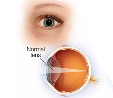
When a person enjoys normal vision without glasses or contacts, light rays enter the eye through the front, clear portion of the eye known as the cornea.
Normal Eye

Normal Eye
When a person enjoys normal vision without glasses or contacts, light rays enter the eye through the front, clear portion of the eye known as the cornea. From there, the light rays pass through the lens of the eye where they are precisely focused on the retina. The retina converts these light images into electrical signals, where they are passed through the optic nerve to the brain. Clear vision results. Like the lens of a camera, if the cornea fails to focus these images properly on the retina, blurred or distorted images can result.
Parts of the Eye

Parts of the Eye
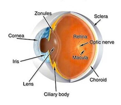
•Cornea: Transparent front part of the eye that covers the iris, pupil, and anterior chamber and provides most of an eye's optical power.
•Iris: Pigmented tissue lying behind the cornea that gives color to the eye (e.g., blue eyes) and controls amount of light entering the eye by varying the size of the pupillary opening.
• Pul: Variable-sized black circular opening in the center of the iris that regulates the amount of light that enters the eye.
• Lens, crystalline ns: The eye's natural lens. Transparent, biconvex intraocular tissue that helps bring rays of light to a focus on the retina. Cataracts usually form in the Lens.
• Reta: Light sensitive nerve tissue in the eye that converts images from the eye's optical system into electrical impulses that are sent along the optic nerve to the brain. Forms a thin membranous lining of the rear two-thirds of the globe.
• Macu: Small central area of the retina surrounding the fovea; area of acute central vision.
• Optic nee: The nerve of the eye; carries impulses for sight from the retina to the brain.
• Vitreous, vitreous huor: Transparent, colorless gelatinous mass that fills the rear two-thirds of the eyeball, between the lens and the retina.